Purpose: To analyze the halo formation of several intraocular lenses (IOLs) in the optical bench.
Setting: University of Murcia, Murcia, Spain.
Design: In vitro study.
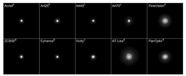
Methods: Light from a green LED passed through a pinhole and was collimated. Each IOL was placed within a realistic model eye having a PMMA cornea with a physiological amount of spherical aberration and a 4.5 mm aperture. A digital camera sensor acted as the retina and a focus tunable lens was used to change the object’s vergence (range ±4 diopters). Series of images were captured with different exposure times and fused to get a high dynamic range image. Performance was assessed by analyzing the corresponding halo brightness and size. The tested lenses, that included biconvex and inverted meniscus IOLs, were monofocals, extended depth-of-focus (EDOF), and diffractive trifocals.
Results: Monofocal lenses produced halos with a radius close to 0.4 degrees. The halo radii of the nondiffractive EDOF lenses ranged between 0.45 and 0.63 degrees, whereas diffractive lenses had radii ranging from 0.84 to 1.22 degrees. The halo was generally dimmer for the refractive lenses and brighter for the diffractive. The through-focus images show that the halo size was larger at any defocus position for the diffractive lenses than for the rest of the tested IOLs.
Conclusions: The diffractive IOLs exhibited a characteristic halo structure. Performance of the inverted meniscus and other nondiffractive lenses (Vivity and Eyhance) was comparable with a monofocal lens. This on-bench test can serve as an indication of the potential impact of photic phenomena on patient satisfaction.